| OBJECTIVE (NUMERICAL APERTURE | RESOLUTION LIMIT (MICROMETERS) | PROJECT SIZE (RESOLUTION X MAGNIFICATION) | CALCULATED REQUIRED PIXEL SIZE (MICROMETERS) |
|---|---|---|---|
| 2X (0.06) | 4.6 | 9.2 | 4.6 |
| 4X (0.10) | 2.8 | 11.2 | 5.6 |
| 10X (0.25) | 1.1 | 11.0 | 5.5 |
| 20X (0.4) | 0.69 | 13.8 | 6.9 |
| 40X (0.65) | 0.42 | 16.8 | 8.4 |
| 50X oil (0.95) | 0.29 | 14.5 | 7.3 |
| 60X (0.80) | 0.34 | 20.4 | 10.2 |
| 100X oil (1.25) | 0.22 | 22.0 | 11.0 |


The Deal with Megapixels
This entry was posted on June 10, 2018.
Is More Better?
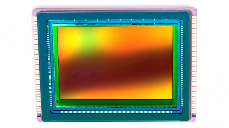
As microscopists and also general consumers, consumer (non-scientific) camera manufacturers have tricked us into thinking “more” megapixels translate into better images. NOT TRUE, at least not in science! Given the same size of the sensor, more pixels (or the tiny little light-sensing component of a camera sensor) means smaller pixels which, in turn, means less “volume” in the pixel to sense light, thus reducing sensitivity. Also, due to the design of the technologies, there is a little more space between pixels in a CMOS sensor than a CCD sensor, therefore CMOS pixels tend to be smaller than those on a CCD.
 Figure 1. Illustration of pixel number and camera resolution.
Another misnomer is that more pixels will give you more resolution. That’s not accurate. The primary determining factor for resolution is the microscope, the Numerical Aperture of the objective, and whether you’ve adequately performed Köhler Alignment. The microscope determines optical resolution, and no camera can “see” more than is already there. Without going into too much detail, “resolution” on a camera simply means that you can tell that 2 points in your specimen are truly distinct features. To confirm resolution, there needs to be at least one empty pixel between the two points. In a very simplistic example (please don't criticize my artistic skills), Panel 1 in Figure 1 illustrates how 3 points may be seen through the microscope. Camera pixel maps are overlaid to show relative sizes of pixels in a lower "megapixel" camera (Panel 1.A) vs. a higher megapixel camera (Panel 1.B). Panel 2 illustrates what the camera pixels register (complete with relative intensity of the boxes). Note in the 8x8 pixel camera (A and A') that there is no way to tell one point from another – it could be just one bigger object of varying intensity. However, the 12x12 pixel camera (B and B') can resolve 3 objects.
Size Really Does Matter
As hinted to above, there is an optimal number of pixels necessary to resolve 2 points in space. It is based upon a theorem from Harry Nyquist (1928) and simplified to state that the frequency of a digital sample needs to twice that of the analog signal. The analog signal is the specimen we are viewing with magnification. So what does that mean to us? Basically, there needs to be an empty pixel between specimens, and magnification also has an impact. The table shows the optimal pixel size for imaging with a given objective, to maximize the optical resolution of the microscope. Note that these values are based on Achromat objectives – objectives with different resolutions (higher or lower Numerical Aperture) would require smaller or larger pixels. Note that the higher the magnification, the larger the pixels! The take home message here is to use a camera with a pixel size that closely matches the lowest objective magnification you plan to use for taking images.
Table 1. Required Pixel Size for Optimal Image Resolution by Objective Magnification
And when it comes to sensitivity for lower light applications, pixel size does matter. Smaller pixels are less sensitive than larger pixels. For simplicity, imagine a pixel as a drinking glass, and a small glass captures fewer water drops than a bigger glass. So for situations where sensitivity is important (i.e. fluorescence, darkfield, phase contrast), larger pixels (and consequently lower megapixel cameras) are actually preferred.
ACCU-SCOPE and UNITRON offer a variety of cameras to suit many imaging applications. Feel free to ask us to recommend a camera that best suits your needs.
Thanks for reading!

