Microscopes are critical tools in forensic sciences. From determining the cause of death to highlighting minute details in trace evidence, microscopes allow tiny details to make large contributions to law enforcement, medical science, and other fields.
Evidence analysis has come a long way from the proverbial magnifying glass of Sherlock Holmes, and advancements in microscope technology have revolutionized forensic investigations. For example, Benjamin J. Jones notes in Microscopy in Forensic Science that modern investigators have access to much more data than their counterparts of ages past. That's true even when the available evidence seems miniscule in both quantity and size.
The analysis is also more in-depth thanks to advancements in microscope technology. Forensic investigators may be able to pull clues from this trace evidence that point to the type of weapon, identify debris related to the crime, and even link fibers, hair, fragments, or fingerprints to an accused subject.
The Stereo Microscope in Forensic Applications
Stereo microscopes provide 3D images of samples that can help investigators better understand the nature of the trace evidence they're viewing.
The 3D visualization is created by combining the microscope's technology with inherent functions of the human brain. Specifically, two optical paths deliver variations of the same image to each of the eyes by altering the viewing angle slightly. The slight difference in viewing angle between both eyes results in subtle illumination and shading variations. The brain receives this input and interprets it as a single 3D image.
Stereo microscopes offer both binocular (uses two eyes) and stereoscopic (3D) visualization, which means there are two eyepieces requiring both eyes and the resulting image as perceived by the brain is in 3 dimensions. This technology increases depth perception, which supports the examination of a sample's topography. Viewing the same sample through a single lens might cause an investigator to miss a critical pattern, dip, or texture that enhances understanding of the sample.
While stereo microscopes can provide a useful magnification of up to 100x, practical use typically involves observations made with magnification between 4x and 40x.
These types of forensic microscopes also support greater working distances than other alternatives, permitting more room for manipulating and positioning the sample. Some microscopes include features such as illumination options or built-in or attachable cameras and screens, allowing researchers to view samples in varying ways or capture images or videos of samples for documentation or to share with others. Most of these options are available as accessories, too.
Numerous applications in forensics exist for stereo microscopes. They include, but aren't limited to:
- Gunshot residue (GSR) and primer residue examination: Forensic scientists can use stereo microscopes to examine trace evidence ranging from bullet fragments to gunpowder patterns. Tiny elements that are unseen by the human eye can fill in the blanks in forensic puzzles under the magnification of a stereo microscope. For example, when a gun is fired, GSR creates patterns on any objects near the barrel. These patterns can help investigators link crimes to specific guns or shooters or put together a narrative about how a crime played out.
- Document examination: According to the Centurion University of Technology and Management, stereo microscopes are well adapted to examining various document traits, including overwriting, erasures, color, and ink. Investigators might, for example, compare microscopic details regarding pen strokes or the differences between paper or printing ribbons to understand when various documents were created and by whom.
- Insect identification in entomological forensics: This technology can help investigators determine the type and origin of an insect species when natural human observation fails. It may seem easy to determine the difference between a green June beetle and a red ladybug, for instance. However, if evidence includes only part of the bug or harder-to-identify eggs or larva, entomological forensics can put the puzzle together with help from stereo microscopes. Insect identification can help investigators understand the origin of evidence or what might have happened to it.
Metallurgical or Polarized Light Microscopes: Tracing the Evidence
Standard microscopes, including stereo microscopes, work with evidence that has been prepared to provide contrast whether the sample is essentially either transparent or opaque. An opaque sample is generally illuminated from above or from an angle. If a sample is thin enough and is illuminated from below, some light may pass through and reveal internal details specific to the sample and its source.
However, this approach doesn't work for all samples — some samples are too thick for light to pass through. A metallurgical microscope lights the sample from above, allowing for viewing and data gathering. The illumination is created in a way to minimize or eliminate glare and shadows that would distract the investigator or interfere with a complete understanding of the sample.
In some cases, light is passed through filters to change its intensity or polarization, an optical characteristic of different materials. Polarized light microscopy allows investigators to increase contrast or otherwise enhance the ability to study a sample and gather more data about its optical properties. Because polarized light enhances the investigator's ability to view minute details of a sample including clues into its chemical composition, it increases accuracy in analyzing trace evidence.
The ability to drill down into details on evidence based on optical characteristics creates many applications for metallurgic microscopes and polarized light microscopy. Some potential applications in forensics include:
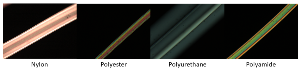
- Examining hairs or fibers: Polarized light microscopy allows researchers to gather helpful details of a case, even if they're working with a single fiber of cloth or a hair. They can determine facts such as whether trace evidence is a natural fiber, synthetic fiber, the composition of the fiber, or hair from a human or animal. Other details they can gather include color, shape, what species the hair belongs to, whether the fiber or hair was cut or torn, and whether dye or other altering materials are present.
- Analyzing soil or minerals: This technology allows researchers to build vast banks of data about soil and minerals from across the globe. For example, the Natural Resources Conservation Service‐Kellogg Soil Survey Laboratory has a database detailing grain counts associated with mineral samples from more than 7,500 sites in the United States. Access to this type of data and the ability to examine trace soil and mineral evidence found at crime scenes or on other pieces of evidence are critical in forensics. They make it possible for forensic investigators to pinpoint locations or create theories about how the evidence came to be in a certain place.
- Determining refractive indices of glass particles: As polarized light passes through samples, researchers can measure the refractive index — how much the light bends as it travels through the material. That measurement is helpful in determining physical characteristics of a sample, such as thickness and purity, which may be important in forensic analysis.
The Unparalleled Precision of the Comparison Forensic Microscope (CFM)
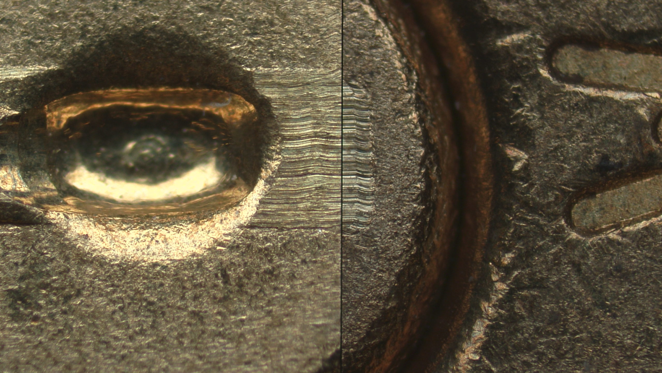
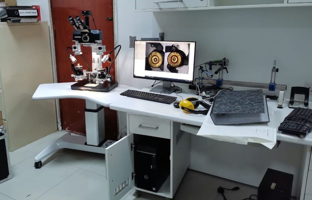
The comparison forensic microscope has become an essential tool in forensic criminal investigations and other research because CFM technology presents side-by-side images of two different samples for simultaneous viewing. It achieves this via a unique split-image feature, the hallmark of the CFM.
Samples are placed on two stages and viewed using two separate microscope instruments, one for each stage. These are bridged up to a single viewing element or output to a single screen, allowing researchers or investigators to see both images at the same time.
The ability to drill down into differences or similarities between two samples is obviously critical to modern forensics, and the CFM offers unparalleled precision at the moment. Forensic scientists can quickly identify important factors of interest between the samples, and this has been especially pivotal in bullet and cartridge case examinations. In one view, criminal forensic investigators can see striations and other markings that help them understand answers to questions such as whether two different bullets were fired from the same gun.
In addition to matching bullets to firearms, other notable uses of CFMs in forensics include tool mark identification (e.g., markings left on a shell casing from the gun hammer) and document examination. For example, this type of microscope can be used to compare known handwriting samples to questioned samples or evidence gathered at a crime scene.
What Does the Future Hold for Forensic Microscopes?
Forensic microscopes are an integration of tried-and-true science and always-evolving technology. In the past few decades, innovations in microscope technology have transformed forensic science, allowing investigators and researchers to derive increasing amounts of accurate data from even the smallest samples.
Continuous innovation allows microscope technology to keep pace with the changing needs and demands of forensic investigations. And evolving technology will allow investigators to answer more questions, answer them faster, and answer them with greater confidence as they examine trace evidence.
UNITRON offers a wide selection of stereo microscopes, illumination solutions, digital imaging solutions, metallurgical microscopes with polarized light capability, and our renown CFM comparison forensic microscope.