Detectives are sorting through piles of evidence when a faint toolmark on a spent cartridge case emerges as the crucial detail in a high-profile case. Traditional forensic techniques can struggle to capture such subtle features, but comparative microscopy brings this microscopic evidence into sharp focus. In an environment where every detail matters, the stakes couldn’t be higher.
The Hidden Clues of Comparative Microscopy
In forensic investigations, even the smallest details can untangle complex questions of cause and responsibility. Minute surface marks—often invisible to the naked eye—can carry critical information about how a crime was committed and what tools were involved. Without the right analytical approach, these clues risk being overlooked or misunderstood.
Comparative microscopy changes that equation. By enabling direct, side-by-side examination of physical evidence, this technique allows forensic scientists to identify similarities and differences with clarity and confidence. What once appeared insignificant can become a defining piece of the investigative puzzle, helping examiners connect evidence to specific actions, tools, or firearms. In modern forensic science, this capability is not just helpful—it’s essential.
Understanding Comparative Microscopy
Comparative microscopy is a forensic technique that allows two or more specimens to be examined simultaneously under matched optical conditions. Unlike traditional microscopy, which focuses on a single sample at a time, comparative microscopy emphasizes direct visual comparison, making it easier to identify corresponding features or meaningful differences.
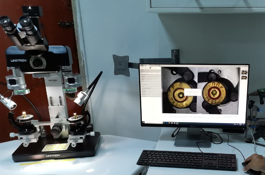
This approach relies on specialized instruments designed for precision and stability. Forensic comparison microscopes use a dual optical path to present specimens side by side within a single field of view. High-quality optics, consistent illumination, and digital imaging capabilities allow forensic examiners to evaluate microscopic details with accuracy and repeatability—critical requirements in firearm and toolmark examination.
Applications in Forensic Science
Comparative microscopy plays a central role in several areas of forensic evidence analysis, including:
- Paint Analysis: Comparing paint layers and surface characteristics to determine a common source.
- Gunshot Residue: Evaluating particulate evidence associated with firearm discharge.
- Toolmark Examination: Analyzing marks left when a tool or firearm component comes into contact with another surface.
- Firearm and Ballistic Evidence: Comparing bullets and cartridge cases to assess whether they were fired from the same weapon.
Toolmark Examination in Practice
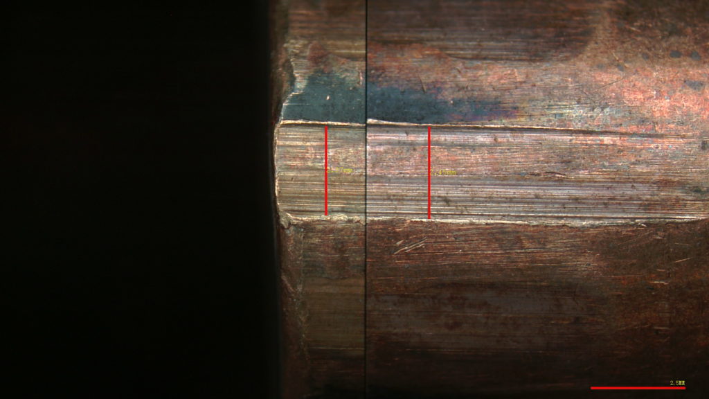
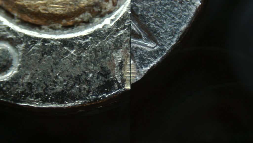
In a hypothetical firearms investigation, forensic examiners use a forensic comparison microscope to analyze toolmarks on a spent cartridge case recovered from a crime scene. By comparing breech face impressions, firing pin marks, and other cartridge case features to test-fired samples from a suspect firearm, examiners can assess microscopic similarities created during the firing process.
This side-by-side analysis allows investigators to determine whether the marks share a common origin, helping establish a reliable association between the evidence and a specific firearm. Such comparisons are a cornerstone of firearm and toolmark examination and demonstrate the critical role comparative microscopy plays in linking physical evidence to real-world events.
Advantages Over Traditional Methods
Comparative microscopy offers several advantages over traditional, single-sample examination techniques:
- Direct visual comparison: Viewing evidence simultaneously reduces interpretation gaps and supports objective analysis.
- High-resolution imaging: Advanced optics and digital documentation capture minute details for review and reporting.
- Improved reliability: Consistent viewing conditions strengthen examiner confidence and support defensible conclusions.
By minimizing subjectivity and enhancing clarity, comparative microscopy improves the overall quality of forensic evidence analysis—particularly in cases involving ballistic and toolmark evidence.
Challenges and Practical Considerations
Like any advanced forensic technique, comparative microscopy requires specialized training and experience. Examiners must understand how toolmarks are created, how to prepare and orient samples, and how to interpret similarities responsibly.
Cost can also be a consideration for some laboratories, as forensic comparison microscopes are precision instruments. However, reliable equipment, technical support, and application-specific design help forensic professionals maximize the value of these systems in daily casework.
The Future of Comparative Microscopy in Forensics
As forensic laboratories continue to modernize, comparative microscopy remains a foundational technique. Ongoing advancements in digital imaging, documentation software, and workflow integration are enhancing how evidence is captured, reviewed, and presented.
Emerging technologies—such as AI-assisted pattern recognition and improved digital comparison tools—are expected to support examiners without replacing expert judgment. As these developments evolve, comparative microscopy will continue to play a vital role in firearm and toolmark examination, supporting accurate, transparent, and reproducible forensic analysis.
The Future of Forensic Precision
In the complex world of forensic science, comparative microscopy stands out as a powerful tool for uncovering truth at the microscopic level. By enabling detailed, side-by-side comparison of ballistic and toolmark evidence, it helps forensic professionals transform subtle markings into meaningful conclusions.
As investigative demands increase and standards for accuracy rise, comparative microscopy remains essential to modern forensic practice. When justice depends on the smallest details, having the right tools—and the ability to interpret them with confidence—makes all the difference.